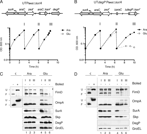
Fig. 2.
FimD folding in conditional E. coli surA mutants. (A and B) Growth curves of static cultures of UTPBAD::surA and UTdegP-PBAD::surA strains grown in rich BHI medium containing d-glucose (Glu) or l-arabinose (Ara) and maintained in exponential phase by repeated dilutions with the same medium. Samples from these cultures were taken at the indicated times (I, II, and III) for Western blot analysis. A schematic drawing of the relevant genetic structure of these strains is depicted at the top of the growth curves. (C and D) Whole-cell protein extracts from bacteria harvested at the indicated times (I, II, and III) from cultures shown in panel A or B were analyzed by Western blotting with r anti-FimD, anti-OmpA, anti-SurA, anti-Skp, anti-DegP, and anti-GroEL antibodies. Samples from UTPBAD::surA are shown in panel C. Samples from UTdegP-PBAD::surA are shown in panel D. Whole-cell protein samples were prepared as indicated in the legend of Fig. 1. Control samples were obtained from cultures of the corresponding strain (UTPBAD::surA in panel C; UTdegP-PBAD::surA in panel D) grown in medium with l-arabinose.

