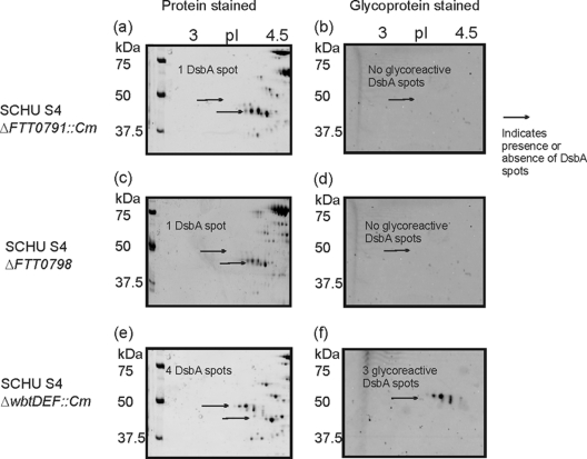
Fig. 3.
2D electrophoretic analysis of F. tularensis subsp. tularensis SchuS4 ΔFTT0791::Cm, ΔFTT0798, and ΔwbtDEF::Cm mutants. Zoom images of DsbA protein resolved by 2D-PAGE. Cell lysates (100 μg) were separated in the pH range 4 to 7. Each gel was stained sequentially with Emerald Q glycostain to visualize glycoreactive protein spots and then with Sypro Ruby protein stain. (a) SchuS4 ΔFTT0791::Cm mutant stained with Sypro Ruby; (b) SchuS4 ΔFTT0791::Cm mutant stained with Emerald Q glycostain; (c) SchuS4 ΔFTT0798 mutant stained with Sypro Ruby; (d) SchuS4 ΔFTT0798 mutant stained with Emerald Q glycostain; (e) SchuS4 ΔwbtDEF::Cm mutant stained with Sypro Ruby; (f) SchuS4 ΔwbtDEF::Cm mutant stained with Emerald Q glycostain.

