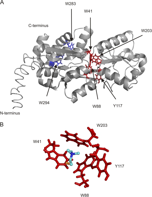
Fig. 7.
Homology model of A. tumefaciens ChoX. (A) ChoX model was generated using the online server I-TASSER (41) and visualized by PyMOL (http://www.pymol.org). The fold is characterized by a bilobal organization into two lobes connected via a linker region. The proposed ligand-binding site is located within the two lobes. Proposed choline-binding residues are shown in red, and two aromatic residues (W283 and W294) outside the binding pocket, depicted as negative controls, are shown in blue. (B) Detailed view of the ligand-binding site of A. tumefaciens ChoX. Proposed residues participating in ligand binding and the ligand choline are shown in stick and in ball-and-stick representation, respectively.

