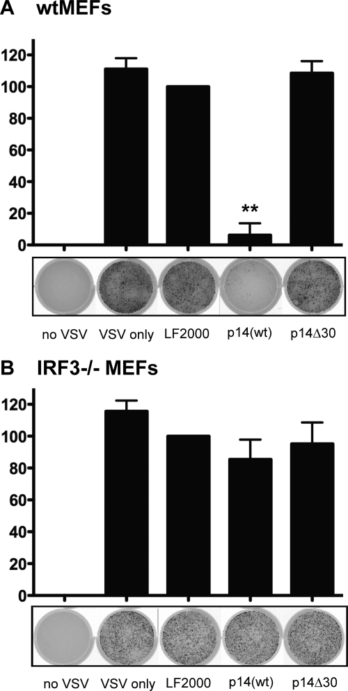
Fig. 3.
p14(wt) triggers an antiviral state that is dependent on IRF3. Following a 24-h treatment with 4 μg of purified p14(wt) or p14Δ30 protein, wild-type (wt) (A) and IRF3−/− (B) MEFs were infected with VSV expressing GFP from the viral promoter. GFP fluorescence was detected 24 h postinfection using a Typhoon Trio imager (GE Healthcare). The level of GFP expression was quantified using ImageQuant TL software (GE Healthcare) and expressed as a percentage of fluorescence relative to that of LF2000-treated wells. Data are presented as means ± standard errors of the means (SEM) from three independent experiments. Statistical analysis was performed by one-way analysis of variance (ANOVA) and Tukey's post hoc test, comparing all treatments to that with LF2000 alone. Cells incubated with no VSV or with VSV only were included as controls. **, P > 0.001.

