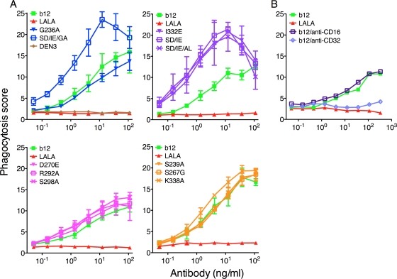
Fig. 5.
Phagocytosis of gp120-coated beads with wt b12, b12 variants, and DEN3. (A) Fluorescent gp120-coated beads were opsonized with antibodies for 2 h before the addition of THP-1 cells. Phagocytosis was evaluated after 24 h of coincubation of cells and bead-antibody complexes using flow cytometry. A phagocytosis score was calculated by multiplying the percentage of cells positive for beads with the mean fluorescence intensity of the same cell population. Applying both values ensures that the number of active phagocytic cells as well as the phagocytic efficiency of the individual cell is added to the experimental read-out. FcγRIIa up-variants are shown in blue, FcγRIIa down-variants are shown in pink, FcγRIIIa up-variants are shown in purple, and FcγRIIIa down-variants are shown in yellow. Values are means and standard deviations of triplicate wells. The assay was repeated twice. (B) As in panel A, except that an anti-CD16 or anti-CD32 antibody was added together with wt b12 to determine the FcγR (IIa or IIIa) that mediated phagocytosis of the beads. The assay was performed twice with similar results.

