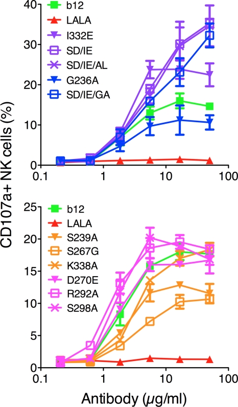
Fig. 6.
In vitro NK cell activation using wt b12 and b12 variants. Microtiter plates were coated with antibody. Freshly isolated NK cells were incubated for 4 h before evaluation for CD107a expression by flow cytometry. Curves were generated by plotting percent NK cell expression as a function of coating antibody concentration. FcγRIIa up-variants are shown in blue, FcγRIIa down-variants are shown in pink, FcγRIIIa up-variants are shown in purple, and FcγRIIIa down-variants are shown in yellow. Values are means and standard deviations of triplicate wells. The assay was performed twice with similar results.

