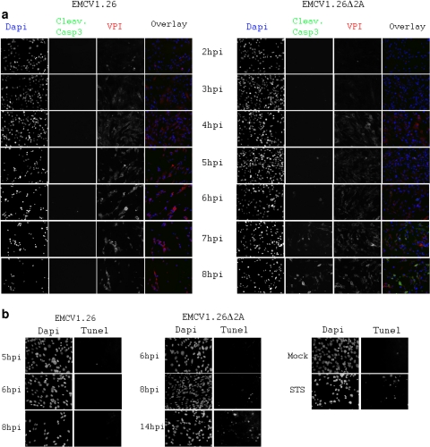
Fig. 10.
EMCV1.26Δ2A-infected cells die by apoptosis, in contrast to EMCV1.26-infected cells. BHK-21 cells were infected at an MOI of 10 with EMCV1.26 or EMCV1.26Δ2A. (a, b) Cells were fixed at the indicated time postinfection and immunostained with anti-EMCV VP1 followed by Alexa 546-labeled anti-mouse IgG (red), with anti-cleaved caspase 3 followed by Alexa 488-labeled anti-rabbit IgG (green), and with DAPI (blue) (a) or labeled for DNA fragmentation using TUNEL staining (Promega) and DAPI (b).

