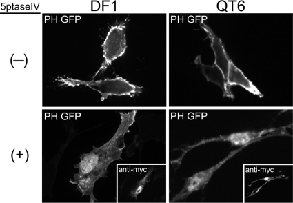
Fig. 2.
Effect of 5ptase on PM localization of PH-GFP in avian cells. DF1 (left) or QT6 (right) cells were cotransfected with DNAs encoding PH-GFP plus a control plasmid (indicated by a minus sign, top) or myc-tagged 5ptase (indicated by a plus sign, bottom). Expression of 5ptase was detected by a monoclonal mouse anti-myc antibody (insets showing the same cells). Images are single, 0.1-μm confocal sections through the midbody of the cell and are representative for each condition (n > 30 cells).

