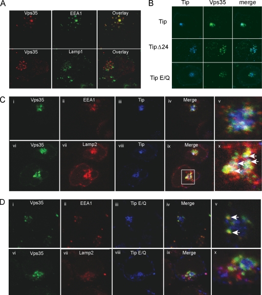
Fig. 4.
Vps35 redistributes to lysosomal compartments in the presence of Tip. (A) Immunofluorescence confocal microscopy of Jurkat T cells expressing Tip or its mutants (blue) and Vps35 (green). (B) Jurkat T cells transiently expressing Vps35 (red) costained with the early endosomal marker, EEA1 (green; top), or with the lysosomal marker, Lamp1 (green; bottom). (C and D) Triple staining of Jurkat T cells for Vps35 (green), EEA1 or Lamp2 (red), and wt Tip or the Tip E/Q mutant (blue). Zoomed images of merged images from the stained cell are shown in the far right column. White arrows indicate the areas of colocalization.

