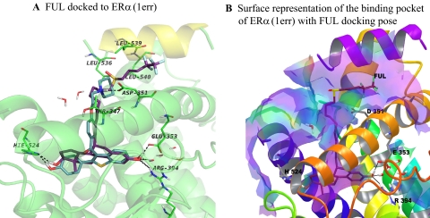
Fig. 7.
Simplified representations of the ERα binding site with fulvestrant. A, representation of the ERα binding site with the best docking pose for fulvestrant (FUL, purple sticks). B, surface representation of ERα binding site accommodating FUL. Hydrophobic areas are mapped in purple, whereas the hydrophilic parts are colored in light yellow-green. The binding site accommodates very well the ligand, which forms the H-bond contacts with the same amino acids like E2 or RAL, whereas the aliphatic side chain protrudes from the binding site and lies in the groove between helix 3 (orange cartoon) and helix 12 (purple cartoon). Only the key amino acids underlying the binding site are shown.

