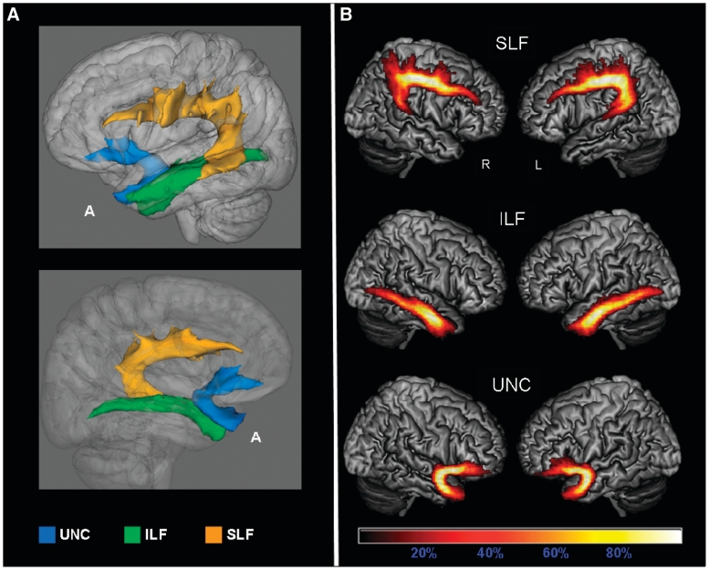
Figure 3.
Probabilistic maps of the language-related tracts from all the subjects included in the study. The tracts are overlaid on a 3D rendering of the MNI standard brain. Only voxels present in at least 10% of the subjects are shown. (A) 3D reconstruction of all-subjects probability maps of left superior longitudinal fasciculus (SLF), inferior longitudinal fasciculus (ILF) and uncinate fasciculus (UNC) seen from left (top) and right (bottom). (B) All-subjects probability maps of bilateral SLF, inferior longitudinal fasciculus and uncinate fasciculus. The colour scale indicates the degree of overlap among subjects. A = anterior.

