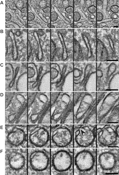
FIG 5 .
Tomographic xy slices through different structures possibly involved in the transition from a tubule to a closed DMV (see also Movie S3 in the supplemental material). (A) A longitudinally oriented tubule reaches its maximum width in the middle panel and fades in the first and last slices. (B) The walls of the tubule have become closer, flattening the compartment and extending it in the z direction, thereby forming a cisterna. Closer membrane pairing can be observed in long stretches of the cisternae shown in panels C and D. (E) Slices through a rounded-up cisterna, essentially an open DMV in a vase-like configuration with an opening to the cytosol (black arrowhead). Fusion of the ends of an open DMV could then give rise to a closed DMV (F). All the panels are 5-nm-thick slices separated by an interval of 15 nm in their respective tomograms; thereby, the encompassed z height is identical for all rows. Scale bars, 100 nm.

