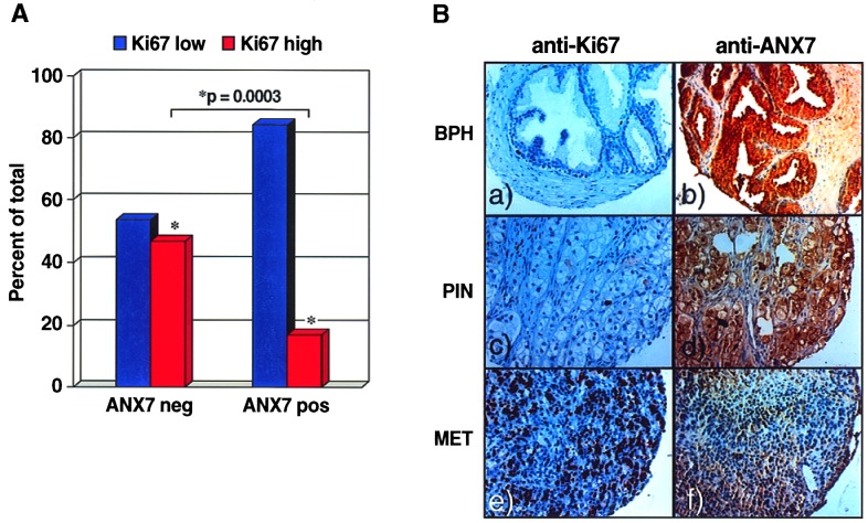
Figure 3.
Levels of ANX7 protein expression and tumor cell proliferation. (A) Relationship between ANX7 expression and Ki67 tumor growth fraction. (B) Representative sections of immunohistochemical staining of Ki67 (Left) and ANX7 (Right) on the prostate tissue microarray (original magnification: ×200). (a and b) Benign prostatic hyperplasia (BPH). (c and d) Primary untreated prostate cancer with only a few scattered Ki67 positive nuclei, but strong ANX7 immunoreactivity. PIN, prostatic intraepithelial neoplasias. (e and f) Distant metastasis (MET) with a high fraction of Ki67-positive tumor cell nuclei and lack of ANX7 immunostaining.

