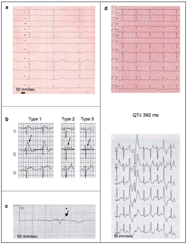
Figure 1.
Long QT syndrome, type 1: prolonged QTc (485 ms), broad-based T waves.
Brugada type 1, 2, and 3 ECGs: Type 1 ECG: Coved-type ST elevation ≥2 mm with T-wave inversion in at least two of leads V1 to V3.Type 2 ECG: Saddle-back ST elevation with increased descent ≥2 mm and at least 1 mm, elevation of the ST segment. Type 3 ECG: Saddle-back or coved-type ST elevation <1 mm.
Epsilon potential in a patient with arrhythmogenic right-ventricular cardiomyopathy
Catecholaminergic polymorphous ventricular tachycardia. Above: Normal resting ECG. Below: Frequent ventricular extrasystoles during exercise

