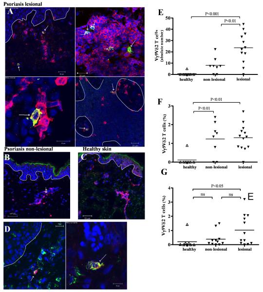
Figure 4. Vγ9Vδ2 T cells are increased in skin of psoriasis patients.
Frozen skin sections were stained with anti-CD3 mAb (red) and anti-Vδ2 mAb (green), identifying Vδ2 T cells as yellow. Nuclei were stained with To-Pro-3 (blue) and dermo-epidermal barrier is indicated with a white line. Vδ2 T cells were present in lesional psoriatic dermis as well as scattered in the epidermis (A). Vδ2 T cells were found in non-lesional psoriatic skin (B) while in healthy skin, Vδ2 T cells were rare (C). Co-staining of anti-CLA (green) and anti-Vδ2 (red) revealed CLA expression on skin Vγ9Vδ2 (D). Psoriatic skin harbored significantly higher absolute numbers of Vδ2 T cells than non-lesional (p<0.01) or healthy skin (p<0.001) as calculated in 1 cm of skin section (E). Relative quantification of Vγ9Vδ2 T cells in skin sections revealed that a higher percentage of T cells in non-lesional (p<0.01) as well as lesional (p<0.01) psoriatic skin express Vδ2 than in normal skin (F). We established T cell lines from lesional (n=12), edge (n=11) and healthy (n=8) skin by culturing skin pieces in the presence of IL-2 (60 IU/ml) and IL-15 (12 ng/ml) for 2-3 weeks and harvesting cells that migrated out of the tissue. Skin T cell lines were analyzed for γδ T cell subsets after gating on CD3+ cells. In lesional skin there were more Vγ9Vδ2 T cells (1.03% (±0.31%)) than in edge (0.39% (±0.16%), ns) or healthy skin (0.21% (±0.18 %), p<0.05) (G).

