
Figure 1.
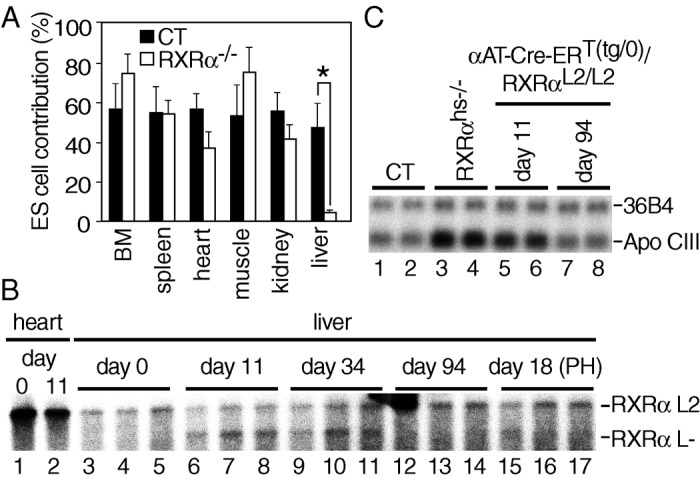
Impaired lifespan of RXRα null hepatocytes. (A) Low contribution of RXRα−/− cells in liver of chimeric mice. The fraction of RXRα−/− cells was evaluated in various organs of 10-month-old chimeric mice by the ratio of the distinct GPI isozymes characteristic of 129/SV and C57BL/6 mice. Six and nine chimeras obtained by injection of CT (RXRα+/+ or RXRα+/− 129/SV ES cells) and RXRα−/− 129/SV ES cells into C57BL/6 blastocysts, respectively, were analyzed. BM, bone marrow. Values are expressed as the mean ± SEM. *, P < 0.001. (B) Time-dependent loss of RXRα L− alleles in liver of Tam-treated αAT-Cre-ERT(tg/0)/RXRαL2/L2 mice. Cre-ERT-mediated RXRα ablation was determined by Southern blot analysis, performed on genomic DNA isolated from heart and liver of αAT-Cre-ERT(tg/0)/RXRαL2/L2 double-transgenic mice, taken before (day 0; lanes 1 and 3–5), and at the indicated days after the first Tam injection (3-month-old animals) (lanes 2 and 6–17). PH was performed 12 days after the first Tam injection (lanes 15–17). Liver DNA was extracted from three animals for each time point. The position of the fragments corresponding to the RXRα L2 and L− alleles is indicated. (C) Relief of down-regulation of ApoCIII expression in liver of RXRαhs−/− and Tam-treated αAT-Cre- ERT(tg/0)/RXRαL2/L2 mice. ApoCIII RNA levels were analyzed by Northern blot, performed on RNA extracted from livers of CT (Tam-treated αAT- Cre-ERT(0/0)/RXRαL2/L2 double-transgenic mouse or vehicle-treated αAT- Cre-ERT(tg/0)/RXRαL2/L2 double-transgenic mouse, lanes 1 and 2, respectively), RXRαhs−/− (lanes 3 and 4), and Tam-treated αAT-Cre-ERT(tg/0)/RXRαL2/L2 double-transgenic mice taken 11 (lanes 5 and 6) and 94 days (lanes 7 and 8) after the first Tam injection. The position of ApoCIII and 36B4 transcripts is indicated.