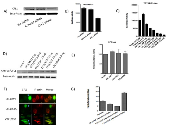
Figure 2.

CFL1 regulates retinoid receptor function. A and B) Jurkat cells were transfected with 2.5 ug of TKRARE-Luc plasmid either in the presence or absence of 2.5 uM control siRNA or CFL1-specific siRNA. Cells were harvested after 36 h and lysates subject to Western blotting using antibodies to CFL1 and beta-actin (A) and luciferase activity measurement (B) as described in Experimental Procedures. C and D) Jurkat cells were transfected with 5.0 ug TATADR1-Luc in the presence of 2.5 ug and 5.0 ug of indicated plasmids. Cells were harvested after 36 h and luciferase activity measured as described in Experimental Procedures (C). Lysates were also subject o Western blotting using antibodies to anti-V5 tag (1:2,000) and beta-actin (1:10,000) (D). E) Jurkat cells were transfected with 5.0 ug of SP1-Luc plasmid in the presence of 5 ug of indicated plasmids. Cells were harvested after 36 h and luciferase activity measured as described in Experimental Procedures. SP1-Luc activity obtained in the presence of vector was normalized to 100%. F and G) Jurkat cells were transfected with CFL1/WT, CFL1/S3A, and CFL1/S3E plasmids and stained with anti-V5 Tag antibodies followed by treatment with F-actin stain as per the instructions of the manufacturer. The cells were visualized using immunofluorescent microscopy (F) and the densitometric mean intensity of F-actin from V5-CFL1 positive cells was quantified and compared to non-V5-CFL1 expressing control cells (G) using AxioVision software from Zeiss Imager D1 fluorescent microscope. Bar, 2 um.
