
Figure 2.
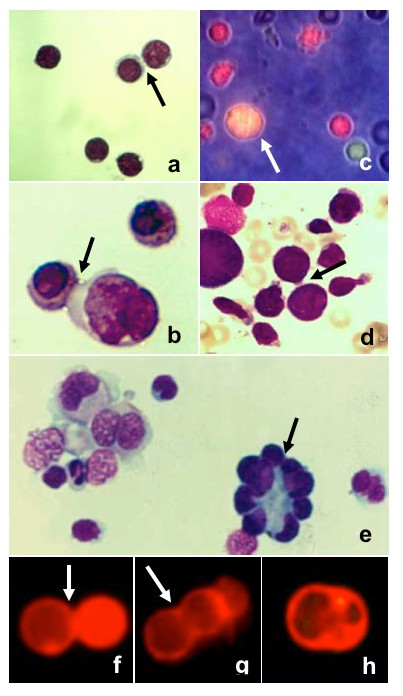
Cells after the first PEG undertreatment (May-Grünwald Giemsa staining). Intercellular bridges (black arrows) were clearly visible: (a) lymphocytes (400×), (b) erythroblasts (800×). One fusion product (white arrow) was present (c) among single cells (green and red), as observed by fluorescent and phase contrast microscopy (500×). (d) Intercellular bridges between erythroblasts (arrow) in a bone marrow smear from a case of erythroleukemia (500×). (e) Single cell suspension from a lymph node affected by Hodgkin's disease (500×). (f) Membrane apposition, (g) triggering of fusion, (h) membrane coalescence, in cells stained by Vybrant DiI (500×).