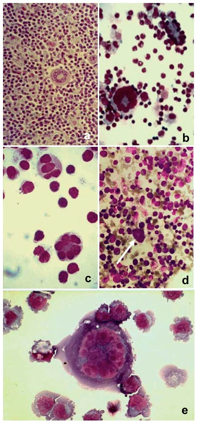
Figure 5.

(a) Histological section of a lymph node from a case of mixed cellularity HD. A Reed-Sternberg cell with nuclei arranged in a circular fashion at the cell periphery is observed in the centre of the picture (200×). (b) Giant polykaryon obtained in vitro when peripheral blood mononuclear cells (PBMC) from an HD patient were co-cultivated with a single cell suspension from the same patient in a Transwell co-culture chamber. The two cell samples are separated by a 0.4-μm microporous membrane permeable to viruses and cytokines. At lower left is a giant polykaryon, and at upper right are several cells in the process of fusing (400×). (c) Giant polykaryon observed after two-step PEG treatment (800×). (d) Giant polykaryon (white arrow) in a bone marrow smear from a case of erythroleukemia (200×). (e) Giant polykaryon from PBMC co-cultivated with P3HR-1 subline in a Transwell co-culture chamber (800×).
