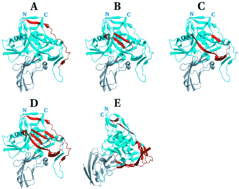
Figure 2.
Anatomical location of the hotspots to exposed, nonhelical loops and strands of the env protein. The Th hotspots are highlighted (red) on the crystal structure (34, 37) of gp120 (blue) complexed with CD4 (gray). The structure can be oriented by the position of the N- and C-terminal residues of gp120 and the CD4 binding site. The regions A–C in this figure correspond to A–C in Fig. 1 with the omission of V2 and V3 sequences, absent from the crystal. The composite of all three hotspots is shown from front (D) and side (E) views.

