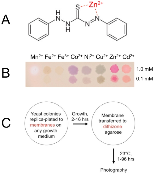
Figure 1. Overview of the dithizone staining procedure.
(A) Chemical structure of dithizone. The zinc ion was placed at the crystallographically determined ligand binding site for zinc [62]. (B) Colored dithizone complexes formed with some transition metal ions. Droplets of 1 mM or 0.1 mM metal sulfate solutions (except chlorides for Co2+ and Ni2+) were applied to a nitrocellulose membrane on a dithizone–DMSO-agarose plate and photographed after one hour at 23°. (C) Schematic of a plate assay for staining yeast colonies.

