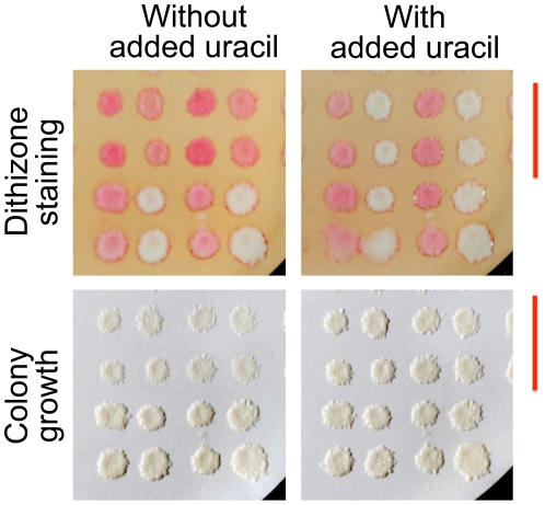
Figure 5. Discovery and correction of partial uracil deficiency in a methionine-poor growth medium.
The Haploid Progeny Collection used in Figure 4 was replica-plated onto a nylon membrane on the medium, without or with added uracil. After overnight growth, the colonies were photographed (lower Panels), stained on dithizone-Triton-agarose plates, and photographed again (upper Panels). For clarity, only the lower right quadrant of the collection is shown (LEU2 HIS3 cells). Image contrast was increased using the same adjustments for the paired uracil conditions. Red bars denote rows of uracil auxotrophs; columns alternate between methionine auxotrophs and prototrophs.

