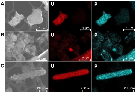
Figure 2. Scanning Electron Micrographs coupled with Energy Dispersive X-ray spectra analysis of Villard ViU-09 soil particles.
For each SEM image, the corresponding EDXS map for uranium (U) and phosphate (P) is presented. (A) soil particles. (B) soil particles and cell-shaped objects. (C) cell-shaped object.

