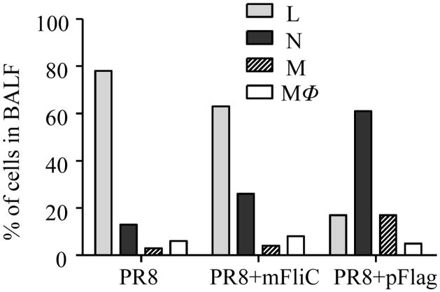
Fig. 8.
Cell populations recruited in BALF fluid. Mice (n=6) were vaccinated with PR8 alone or with mFliC or pFlag and 16 hours later BALF was collected. The cell pellets from BALF of individual mice were resuspended in PBS and smears of them were prepared on slides, air-dried, stained with a modified Wright-Giemsa stain and counted based on cell morphology. Percentages of cell populations were calculated from at least 500 cells counted per optic field in several optic fields. Naïve mice were used as controls (not shown). L: lymphocytes, N: neutrophils, M: monocytes, MΦ: macrophages.

