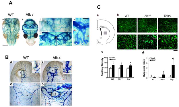
Fig. 2. Brain AVM in Alk1 or Eng deficient mice.
A. Endothelial Alk1 deletion results in AVMs in the brain [38]. A–E. Dissection microscopic views of vascular images of control (WT, A, C) and mutant (Alk1−/−; B, D, E) in postnatal day 3 mouse brains by latex dye injected into the left ventricle of the heart. Magnified views of blood vessels in the hipocampal area (D, E). Asterisks indicate peculiar looping of vessels at the distal tips of arteries shunting to veins (E). A, artery; V, vein. B. Wounding can induce de novo AVM formation in Alk1-deleted adult mice [38]. Vascular patterns shown by latex dye injected into the left heart of control (WT, A, C) and mutant (Alk1−/−, B, D) mice bearing wounds in the ear (A, B) or dorsal skin (C, D), 8 days after induction of Alk1 gene deletion. The images were taken after clearing in organic solvents. Center of the wound is indicated by asterisks. Note that only mutant mice developed AV shunts shown by the presence of latex dye in both arteries and veins. AV shunting and abnormal vascular morphologies were apparent only in the wound areas. Blood vessels away from the wound indicated by arrows with asterisks (B and D) showed normal appearance. Inset in D shows a magnified view of AV fistulas formed in the rim area of the mutant wound. C. Overexpression of VEGF in the striatum of Alk1 and Eng haploinsufficient mice resulted in vascular dysplasia [19]. a. Injection site (grey square). b. Angiogenic foci and dysplastic capillaries (arrows). Inserts are enlarged images of dysplastic capillaries. Scale bars: 100 μm (top panel) and 50 μm (bottom panel). c and d. Capillary density and dysplasia index. * = p<0.05, vs. AAV-LacZ group. # = p<0.05, vs. AAV-VEGF-transduced WT or Alk1+/− mice. VEGF: AAV-VEGF-injected mice; LacZ: AAV-LacZ-injected mice.

