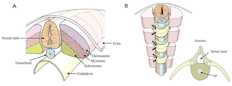
FIGURE 2.
(A) During segmentation the paraxial mesoderm forms pairs of somites along the neural tube (light orange) and notochord (blue). Each somite is composed of a dermatome (light purple), myotome (light brown), and sclerotome (dark yellow). The ectoderm lies above and the endoderm below. (B) Sclerotomal cells migrate from adjacent somites above and below each future vertebra. Dermatomal cells stream beneath the ectoderm to form the dermis, while the myotomal cells form muscle. Insert B shows the architecture of the vertebrae with the spinal canal, spinal processes, and the nucleus pulposus in blue. Courtesy of Dr. Richard Dryden.

