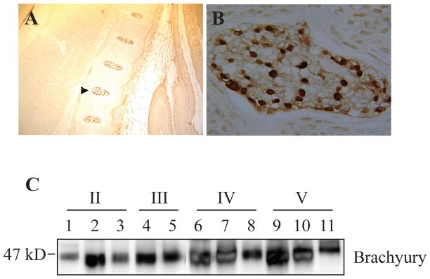
FIGURE 3.
Brachyury expression in cells of the nucleus pulposus. A) Low magnification saggital section of the E15.5 mouse embryo showing brachyury staining in the developing nucleus pulposus. Note that the annulus fibrosus and endplate cartilage is negative (10X). B) High magnification image of the developing nucleus pulposus that is shown by an arrow in panel A (20X). All cells show intense nuclear staining of brachyury. C) Western blot analysis of brachyury expression in nucleus pulposus tissue isolated from progressively degenerate human discs (Thompson Grade II–V). A robust expression of brachyury was seen in all the nucleus pulposus samples. Reproduced with permission.23

