Figure 2.

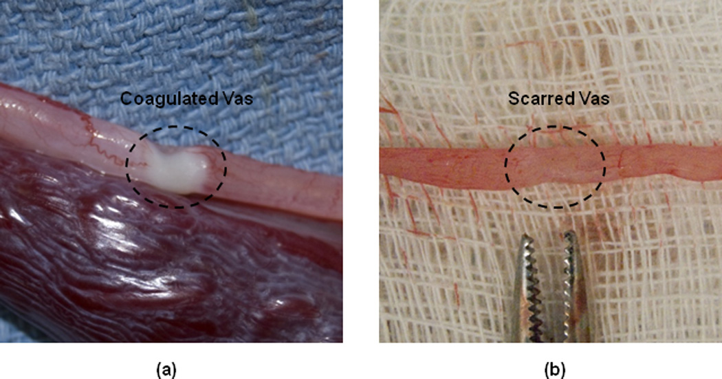
Representative images of the excised canine vas: (a) At Day 0 immediately after the procedure, showing the thermally coagulated zone; and (b) At Day 28, showing the scarred region of the vas.

Representative images of the excised canine vas: (a) At Day 0 immediately after the procedure, showing the thermally coagulated zone; and (b) At Day 28, showing the scarred region of the vas.