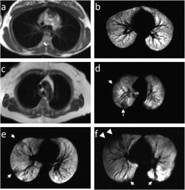
Figure 4. MR of lungs in health and asthma.
Examples of conventional proton MR images paired with corresponding slices from HP He MR images in a healthy normal subject without ventilation defects (a and b) and a healthy normal subject with ventilation defects (c and d, arrows). HP He MR images in a patient with mild-moderate persistent asthma (e) and severe asthma (f). Note the greater central extent of the defects more typical of severe asthma (f, arrowheads).

