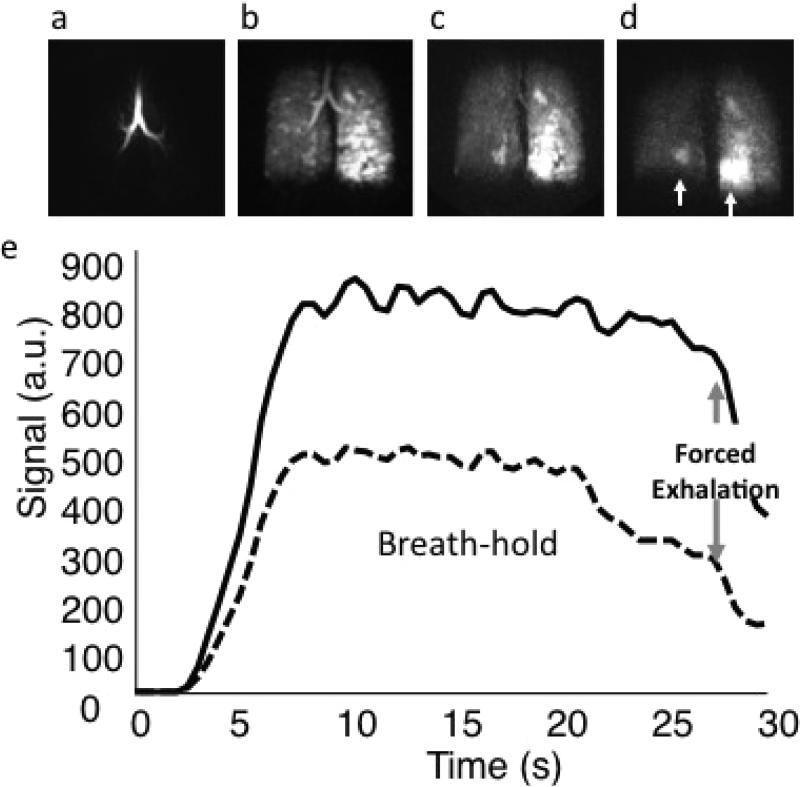
Figure 5. Functional MR in asthma.
Three dimensional dynamic HP He MRI showing gas wash-in (a), breath-hold (b), and forced expiration (c and d) at a 0.5 second frame rate for a patient with moderate persistent asthma demonstrating hetereogeneous gas distribution (b) with gas trapping in the lower right and, most prominently, in the lower left lung (c and d, arrows). (e) Kinetics of wash-in and wash-out derived from regions of interest in the right upper lobe (dotted) and left upper lobe (solid) can be used to quantify regional spirometry.

