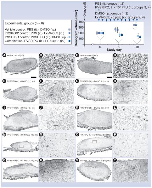
Figure 7. Testing PVSRIPO and PI3K inhibitor synergy in vivo.
Experimental groups and the study regimen are indicated at the top. U-118 xenografts were measured at study days 0 (when PVSRIPO/vehicle and LY294002/vehicle treatment was initiated), 5 and 10. Four animals from each group were euthanized at study days 5 and 10 for histology and virus recovery. The bottom panels show histology from xenografts recovered at day 5 (left columns) and 10 (right columns). Low-magnification images in the left columns are accompanied by higher-magnification images from the same section (inserts) in the column to their right. (A–D) Tumor histology of a representative xenograft from group 1 shows the characteristic dense arrangement of tumor cells. (E–L) Histology of two representative tumors from group 3 at study days 5 and 10 as indicated. Note extensive tumor cell loss and ‘empty’ appearance of the former xenograft in all cases. Arrows point to areas of intense tumor cell killing and active tissue rearrangement. (M–T) Histology of four representative tumors from group 4 at study days 5 and 10 as indicated. (M & N) Complete tumor regression at study day 5. (N & P) The area of the former tumor was invaded by cells with fibroblast morphology surrounded by dense extracellular matrix. Isolated viable tumor cells (arrow [N]) may remain. (Q & R) Active tumor, which was still present in three out of four animals of group 4.
DMSO: Dimethyl sulfoxide; ip.: Intraperitoneal; it.: Intratumoral; PBS: Phosphate-buffered saline; PFU: Plaque-forming unit.
Reproduced with permission from [61].

