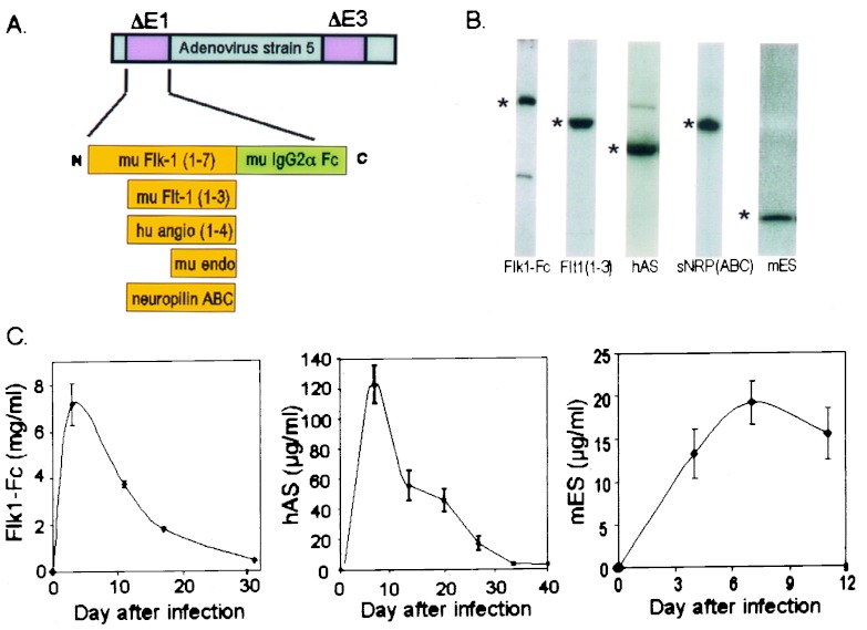
Figure 1.
Construction and characterization of antiangiogenic adenoviruses. (A) Schematic of insertion of various antiangiogenic cDNAs into the E1 region of E3-deleted adenovirus type 5. (B) Western blot analysis of adenovirus-expressed antiangiogenic proteins in mouse plasma. C57BL/6 mice received i.v. injection of 109 particles of the appropriate adenovirus, followed after 2–3 days by Western blot of 1 μl of plasma, except for Flk1-Fc which was taken at day 17 and was a 1:10 dilution. *, position of transgene products: Flk1-Fc (180 kDa), Flt1(1–3) (53 kDa), ES (20 kDa), AS (55 kDa), and sNRP-ABC (120 kDa). Levels in adjacent blots are not comparable because of different enhanced chemiluminescence exposure times. (C) Pharmacokinetics of expression from antiangiogenic adenoviruses. Plasma from mice infected i.v. with 109 pfu of the appropriate adenovirus was analyzed after the indicated times for expression by ELISA (Flk1-Fc, n = 4; ES, n = 4; AS, n = 3). See the text for further details. Error bars, ± 1 SD.

