

Summary
This paper presents various aspects of severe burns involving epileptic patients, who may suffer dramatic accidents during seizure attacks. Epileptics may fall onto an open fire or hot surface (e.g. a kitchen range) and they may upset containers full of boiling liquids, suffering deep burns and scalds. In our experience in this field, the most commonly affected body areas are the face and hands, the trunk, and the lower limbs. All such injuries are full-thickness burns, owing to the very long contact of the skin surface with the lesional agent. Three cases are presented of epileptics with severe burns who were admitted to the Burn Unit of Targu Mures Teaching Hospital, Romania, where they were hospitalized; conservative debridement using polyurethanefoam (PUR-foam) dressings was the standard procedure, which all the patients received. Split-thickness skin grafting was the final method for closing the granulating bed resulting from the conservative debridement. We have found that conservative debridement using PUR-foam dressings is a cheaper and more reliable alternative than sharp debridement (which may remove healthy tissue at the same time as burn eschars).
Keywords: epilepsy, major seizure attacks, full-thickness burns, conservative debridement, PUR-foam dressings, split-thickness skin grafts
Abstract
L'auteur présente les divers aspects des problèmes des patients grands brûlés atteints d'épilepsie, qui peuvent subir des accidents dramatiques au cours de leurs attaques. Les épileptiques peuvent tomber sur les flammes du feu ou sur une surface chaude (par exemple une cuisinière) et ils peuvent renverser des conteneurs pleins de liquides en ébullition, provoquant des brûlures et des ébouillantements profonds. Selon l'expérience de l'auteur, les zones du corps les plus touchées sont le visage et les mains, le tronc et les membres inférieurs. Toutes ces lésions sont des brûlures de toute l'épaisseur de la peau, en raison du très long contact de la surface cutanée avec l'agent lésionnel. L'auteur présente trois cas de patients épileptiques atteints de brûlures graves qui ont été traités dans l'Unité des Brûlés de l'Hôpital Târgu Mures en Roumanie, où ils ont été hospitalisés; la thérapie standard, que tous les patients ont reçue, était le débridement conservateur avec l'emploi de pansements de mousse de polyuréthane. La méthode utilisée pour fermer définitivement le lit de granulation créé par le débridement conservateur a été le greffage à épaisseur variable. L'auteur a constaté que le débridement conservateur avec l'emploi de pansements de mousse de polyuréthane est une alternative moins coûteuse et plus fiable que le débridement agressif (qui peut enlever les tissus sains en même temps que les escarres).
Introduction
Epilepsy is a severe neurological brain function disorder which may begin in early life and leave patients with a severe handicap. Idiopathic epilepsy occurs with no involvement of the brain structure and has two major aspects:
generalized epilepsy (also known as "grand mal") or major seizure, with a characteristic onset and tonic-clonic convulsive movements;
"absence" attack (also known as "petit mal" seizure), which especially affects children and also has a characteristic aspect.
When the brain structure is damaged by various factors (such as trauma and tumours) the seizure attack usually has the form of partial epilepsy (also known as focal or symptomatic) such as temporal lobe epilepsy and the Jacksonian motor seizure.
During seizure attacks patients may suffer severe trauma such as limb fractures, head and neck injuries, and occasionally even deep burns. The most famous description of a seizure attack, with respect to such accidents, is the case of the young epileptic male in the New Testament (Matthew 17:14-15, Luke 9:37-40),1,2 who "often falls into the fire or into the water"…"he suddenly screams, it throws him into convulsions so that he foams at the mouth…."
Most burns in epileptic patients occur during major seizures but some may affect those with "absence" and partial epilepsy, owing to contact with overheated objects. Such burns and scalds are always full-thickness; the patients present a variable total body surface area (TBSA) burned and require admission to a burns unit and appropriate care. The areas of the body most commonly affected are the face and anterior neck, the upper limb, the trunk, and the lower limbs.Three patients with deep burns, suffering from different forms of seizure, are presented in detail in this paper (the term "deep burns" is used in the meaning of full-thickness burns which always require skin grafting). Polyurethanefoam (PUR-foam) dressings (Ligasano) were used for the conservative debridement of all these burn wounds. These synthetic dressings are made of pure white PUR foam, which is highly structured like a honeycomb, which gives the Ligasano sheets (available in thicknesses of 0.5 cm, 1 cm, and 2 cm) the following special properties:
good absorptive power, which enables the Ligasano dressings to drain all scabs, exudate, debris, and pus from all necrotic and infected wounds;
high elasticity, permeability to air, and thermal insulation, preventing wounds from desiccating and serving to cool them;
permanent mechanical stimulation of the wound surface, thus improving the local blood flow with important consequences for natural wound healing;
maintenance of a permanent moist environment (containing active cells, nutrients, growth factors, cytokines, etc.), which is very useful for conservative debridement and wound granulation.
All these qualities listed above - and many others - have persuaded us to use Ligasano dressings for the conservative debridement of full-thickness burns and other acute or chronic wounds, in the last 11 years.
Three case histories
Case 1. Deep scald due to boiling water in the abdomen, external genitalia, and anterior aspect of both thighs
For several years this 55-yr-old male patient, a heavy smoker with significant alcohol addiction, had suffered from short periods of unconsciousness (during which he often fell down). He thought that his symptoms were the result of alcohol and smoking abuse, and he therefore did not ask the family doctor for medical evaluation and treatment.One day, when he was carrying a large pot of boiling water, he had a sudden attack, falling down, overturning the hot liquid onto his abdomen, external genitalia, and thighs, and suffering a deep scald in about 20% TBSA (Fig. 1). Despite the severity of the accident, he came to the Burn Unit only after about three weeks, during which time he was treated at home with silver sulphadiazine dressings by the family GP, an elderly practitioner who did not realize the gravity of the injury.
Fig. 1A. Patent's burns on admission (dark brown adherent dry eschars slightly detaching at edges, with massive staphylococcal infection and characteristic odour).

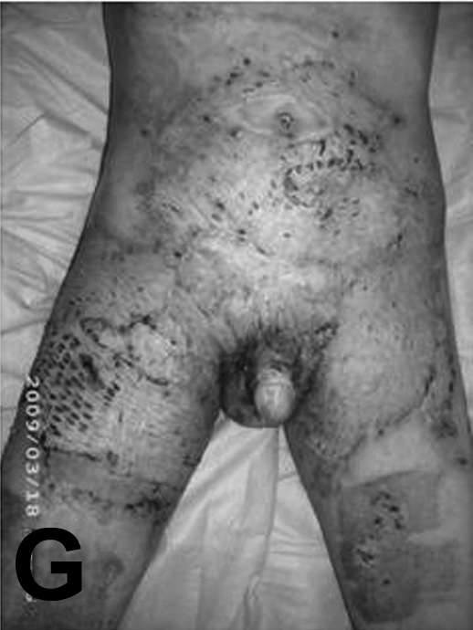
On the day of admission, the burn wound presented dark brown adherent dry eschars, slightly detached at the edges, and there was massive staphylococcal infection with characteristic creamy yellow-grey pus and a specific odour (Fig. 1A). An immediate tangential excision of the superficial infected layers was performed, preserving the adherent deep layers of the burn eschars (i.e. "incomplete tangential excision"), thus avoiding the excessive bleeding that may complicate the regular procedure and at the same time removing the most infected parts of the full-thickness burn wound; the remaining slough was subsequently debrided (conservative debridement) by successive PURfoam dressings (Ligasano) (Fig. 1B). Thanks to these synthetic dressings, all the scabs and infected areas became clean and covered by healthy granulation tissue (Fig. 1C). These granulation areas were eventually covered with meshed split-thickness skin grafts (STSG), harvested with a Watson-Thackray skin-graft knife, and hand-meshed using a no. 20 regular scalpel blade (Fig. 1D). Ten days after grafting, a full take could be seen, as also good spontaneous epithelialization of the donor sites (Fig. 1E). A month after grafting, all the grafts were very well integrated and the donor sites completely healed (Fig. 1F). The patient came once for a follow-up visit, about two months after the skin coverage, but did not appear subsequently for follow-up visits and scar management.
Fig. 1B. Incomplete tangential excision, preserving the adherent deep layer of the burn eschars.
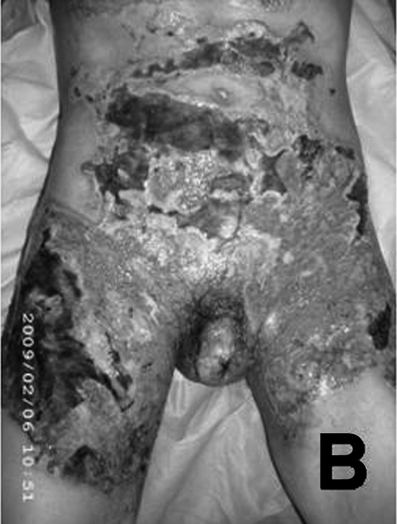
Fig. 1C. Granulating bed following three weeks of PUR foam and topical medication dressings.
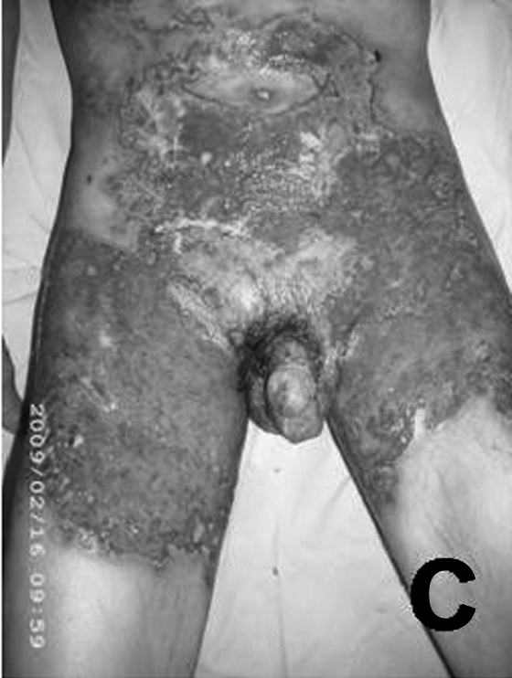
Fig. 1D. Wound bed in operating room before skin grafting (mechanical debridement for granulation tissue).
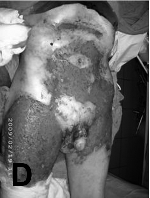
Fig. 1E. Manually meshed autograft in place.
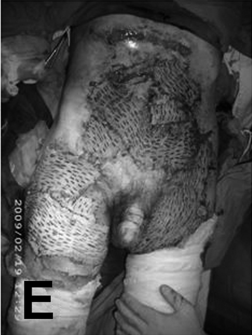
Fig. 1F. Full graft take ten days after grafting.
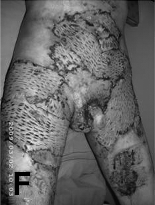
Fig. 1G. Patient’s last visit two months post-graft.
Case 2. Deep upper limb contact burn caused by an overheated metallic surface
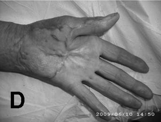
This lesion was suffered by a 73-yr-old patient who suffered periodically from seizure attacks of "absence". During such moments of "petit mal", patients do not have convulsive movements, as in the generalized form of epilepsy, but they are completely "absent" (but not unconscious), immobile, anaesthetic, and underactive; when the seizure stops (the attack can last from seconds to minutes), a period of complete amnesia, disorientation, and sleepiness follows. Our patient had such periodical attacks of "absence", despite a good neurological follow-up and treatment, and during one such seizure she put her left hand on an overheated kitchen broiling surface, suffering fullthickness burns on the volar aspect of the fingers and palm and the lower third of the left forearm (Fig. 2). The initial appearance of the full-thickness contact burn was that of discoloured, dry, thick, waxy eschars covering the volar aspect of the fingers and palm and the lower third of the forearm (Fig. 2A). The patient was unable to say how long she had stayed in that position or specify the circumstances in which the seizure occurred - possibly her daughter had brought her to the burn unit, but she refused to be admitted and operated on. She was initially treated - for about 5-6 weeks - as an out-patient, until the full-thickness burns were debrided (using conservative and autolytic debridement) by alternating the Ligasano PUR-foam dressings and dressings with silver sulphadiazine and PVP-iodine cream and ointment, until a rich granulation bed was obtained (when the patient eventually agreed to be admitted and operated on). The granulation was covered with an unmeshed STSG (Fig. 2B), and the post-operative follow-up after five weeks showed full graft take and good integration as well as an acceptable function corresponding to the patient's age and degree of injury. Despite the fact that the finger burns had not been grafted, very good spontaneous healing occurred (Fig. 2C). The four-month follow-up showed good results and acceptable function, despite the starshaped palmar scar band contracture (the patient refused any possible surgical repair, telling the plastic surgeon she was very happy with the present result) (Fig. 2D). Although contact with an overheated surface is the most frequent cause of deep burns in epileptic patients, there are also other cases when, for example, such patients have dipped their hands into pans containing hot cooking oil, burning stoves, hot driers, and other heated devices, also suffering additional mangling injuries that may lead to the amputation of the fingers and hand and even of the forearm.
Fig. 2A. Initial aspect of the full-thickness contact burn.
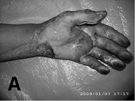
Fig. 2B. Rich granulation in operating room immediately before skin grafting; a skin graft (unmeshed split-thickness skin graft) had already been placed on the lower third of the left forearm.
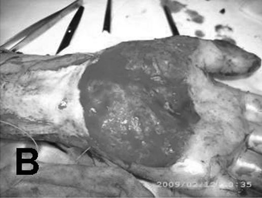
Fig. 2C. Five-week post-operative follow-up: the digital burns were not grafted and healed spontaneously.
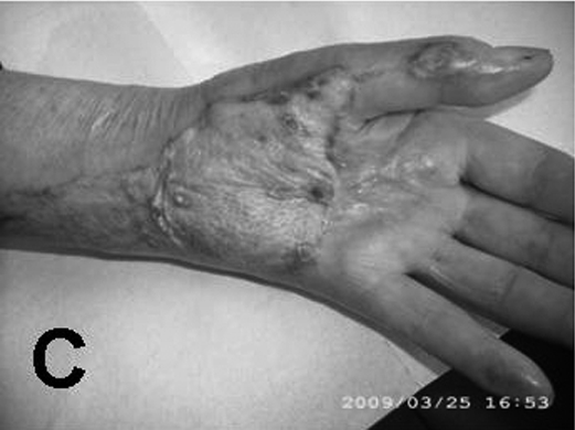
Fig. 2D. Four-month follow-up: nearly full extension despite starshaped palmar contracted scar.
Case 3. Right hemifacial deep burn in a 31-yr-old female patient suffering from frequent generalized seizure attacks
Deep facial and anterior neck burns following contact with hot metallic or ceramic surfaces are severe and devastating lesions with the most dramatic of functional and physiognomical outcomes. Such injuries may affect the palpebral area and the eyeball, with the major consequence of loss of the injured eye. Our statistics of the last 20 years show that over 66% of our epileptic patients with deep facial burns lost the injured eye.
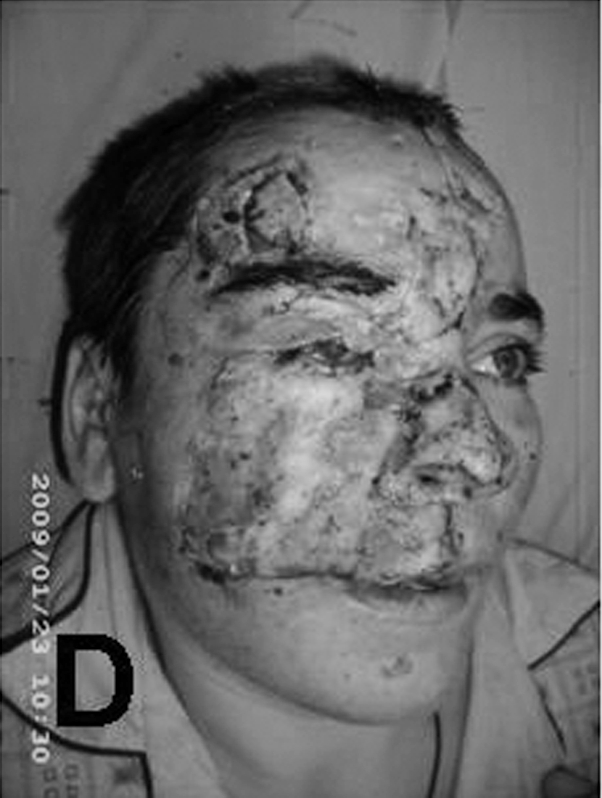
The following case is that of a 31-yr-old female patient suffering from frequent major seizure attacks (2-3 per week) despite continuous neurological medication, details of which are provided later in this paper. During one of her seizures she fell and the right half of her face came into contact with the hot iron door of the heating stove, suffering full-thickness contact burns (Fig. 3). On admission, the initial appearance of the full-thickness contact burn in the right hemifacial area presented dry, waxy, desiccated eschars involving the forehead, nose, eyelids, right cheek, and the right half of the upper lip (Fig. 3A). The burn wound was debrided (autolytic conservative debridement) by alternating PUR-foam dressings with silver sulphadiazine (Fig. 3B), the thick eschars progressively being replaced by a good granulation bed. We do not use routine early sharp debridement for full-thickness facial burns - we prefer conservative autolytic debridement and try to preserve all non-injured tissue. In over 30 years' experience we have found that early complete facial tangential excision of the burn wound may also remove healthy structures, leaving eventually conspicuous ugly scars which are subsequently very difficult to repair. Following conservative debridement, as described above, in about three weeks we obtained a very good granulation bed (Fig. 3C). This bed was covered with unmeshed STSG, which ten days later showed full take, with a few areas of superficial epidermolysis as is often seen under tie-over dressings (Fig. 3D).
Fig. 3A. Initial aspect of full-thickness contact burns in right hemifacial area, presenting dry, waxy, dehydrated eschars involving the forehead, nose, eyelids, right cheek, and the right half of the upper lip.

Fig. 3B. The burn wounds were debrided (autolytic passive debridement) alternating PUR-foam dressings and silver sulphadiazine. In this way the thick eschars were progressively removed and replaced by a good granular bed; we do not normally use sharp debridement for facial full-thickness burns (preferring passive debridement) because all viable healthy tissues have to be carefully preserved. Complete early excision of facial burn wounds may remove healthy structures, leaving conspicuous ugly scars that are very difficult to repair.

Fig. 3C. Following the procedure shown in Fig. 3B, a very good granular bed was obtained in about three weeks.
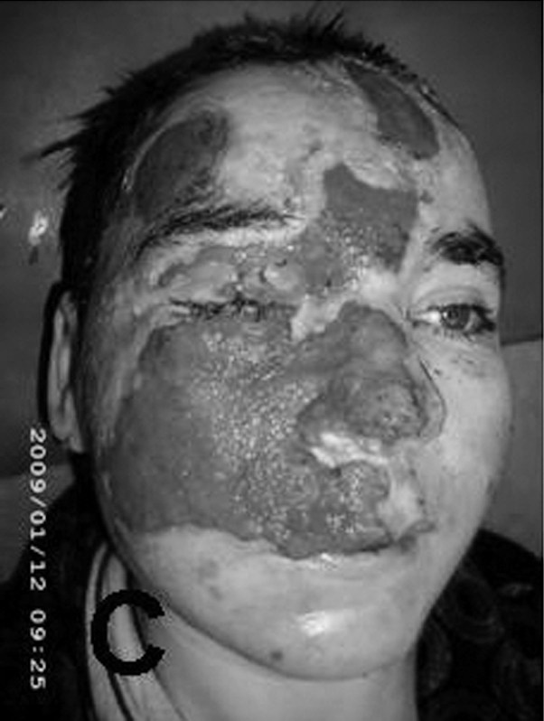
Fig. 3D. The remaining defect shown in Fig. 3C was covered with an unmeshed thick split-thickness skin graft. The photo, taken from above, shows the appearance 8-10 days after grafting (all the grafts took very well, with superficial epidermolysis, which is quite normal under a tie-over dressing).
Six months later the patient presented to our centre with very well integrated skin grafts, but with peri-orbital scars as well as lower eyelid ectropion and internal epicanthi deformation due to the thick contracted scar (Fig. 3E). The inner canthal fold and the scar contracture of the upper eyelid were subsequently released by a Z-plasty and the ectropion of the lower eyelid was excised (Fig. 4), the skin defect being covered with a full-thickness skin graft (FTSG) harvested from the right iliac fossa. We prefer this area as a donor site for FTSG (rather than the "classic" retroauricular skin) because in our experience such grafts retract less and usually provide a long-lasting result (Fig. 4A). This FTSG is then covered by a tie-over dressing which is subsequently removed 7-9 days later; owing to the concomitant Z-plasty (with 45° angles), the ciliary margin of the upper eyelid becomes aligned (Fig. 4B). The follow-up visit after two weeks showed complete palpebral occlusion, normal position and orientation of the eyelashes, and an acceptable dimension of the right eyelid opening (Fig. 4C). A subsequent follow-up visit again confirmed the good aspect of the right eyelid (good occlusion and acceptable symmetry). There is no doubt this patient will need other reconstructive procedures due to the evolution of the scarring process, but at least for the present there are no major physiognomical changes that might have a strong psychosocial impact on her (Figs. 4D and 4E).
Fig. 3E. The photo, taken from above, shows the patient's aspect six months after skin coverage with very well integrated grafts but also presenting palpebral scars (lower eyelid ectropion and internal epicanthi due to the thick scar bands).

Fig. 4A. The inner canthal fold and the scar contracture of the upper eyelid were released by a Z-plasty, the ectropion of the lower eyelid was excised, and the skin defect was covered with full-thickness skin graft (FTSG) harvested from the right iliac fossa; we prefer this area as an FTSG donor site (rather than the classic retroauricular skin) because such grafts retract less and provide a longer lasting result.
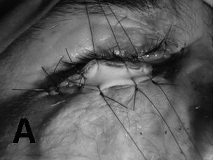
Fig. 4B. The graft shown in Fig. 4A was then covered with a tieover dressing which was removed after 7-9 days; it is possible to see the Z-plasty (with 45° angles) by means of which the ciliary margin of the upper eyelid was aligned.
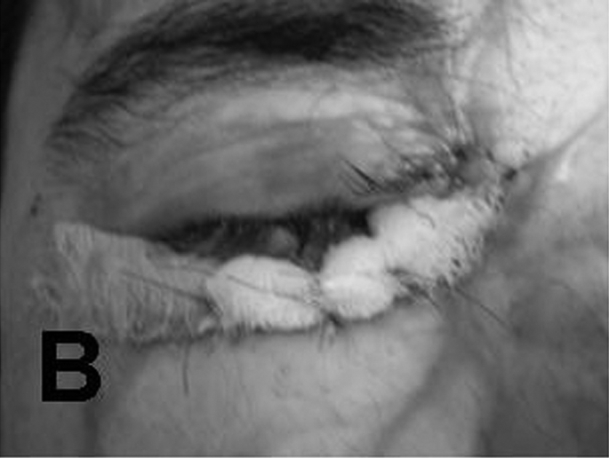
Fig. 4C. Follow-up two weeks post-operation shows complete palpebral occlusion, normal position and orientation of eyelashes, and acceptable dimension of right eyelid opening.
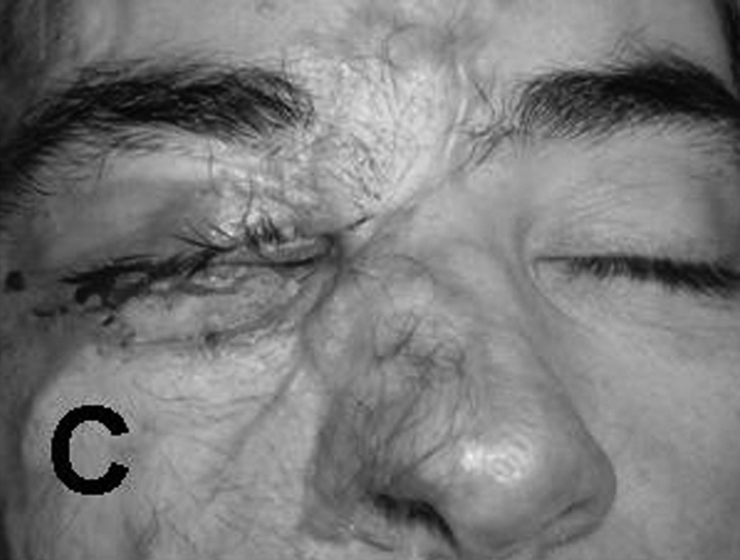
Fig. 4D. The same good result can be seen here after ectropion release and correction of post-burn inner canthal fold.

Fig. 4E. This last figure shows once again the good aspect of the eyelids (good occlusion and acceptable symmetry). There is no doubt this patient will need further reconstructive procedures following the evolution of the scar process, but at least for the moment there are no major physiognomical changes that might have a strong psychosocial impact.

Discussion
In this presentation we have emphasized the severity of burn problems in epileptic patients, despite the fact that such injuries are not very common (10-15% according to statistics over the last 20 years). Facial burns and burns of the upper limb are the most frequent, followed by injuries to the trunk and lower limb. All these burns are full-thickness, owing to the long contact time with the burn agent during seizure attacks. Also, epileptic patients can be difficult patients to treat because often they do not cooperate, refusing lengthy and numerous surgical reconstructive procedures. The association between the neurological disease and the burn injury, combined with the protracted inpatient period in a burns unit and the even longer time necessary for rehabilitation, may lead to deterioration of the basic disease, as epilepsy requires a strong anticonvulsive treatment scheme and a permanent neurological follow-up. For the same reasons, the less traumatic nonsurgical debridement (using autolytic means such as topical medication or PUR-foam dressing) is considered a better option than early excision and grafting (even though the latter substantially shortens the healing and in-patient period). The great risk of general anaesthesia in such patients and the long-lasting neurological effects of anaesthetic drugs in epileptics may discourage surgery in many cases. In facial burns, early excision always removes healthy tissues together with the burn eschars, sometimes with permanent negative effects on the final result.
Conclusion
Many epileptics with facial burns are left with major physiognomical distortion, loss of vision, and other problems, and all these sequelae have enormous psychological, social, and professional consequences. This is the reason why our major concern is to perform a less invasive "stepby step" conservative debridement using the PUR-foam dressings described above (especially for facial burns) and as far as possible avoiding sharp debridement (which always removes health tissue that is extremely useful for better wound repair).
Last but not least, the best way to care for epileptic patients is to prevent them from getting burned in the first place by making impossible all contact with boiling liquids, overheated objects, and other sources of thermal energy, both at work and especially in the home.
References
- 1.Oxford Concise Medical Dictionary (4th edition) Oxford University Press; Oxford: 1994. pp. 223–4. [Google Scholar]
- 2.Holy Bible, New Testament. Vol. 111. Trinitarian Bible Society; London: 2003. pp. 33–31. [Google Scholar]