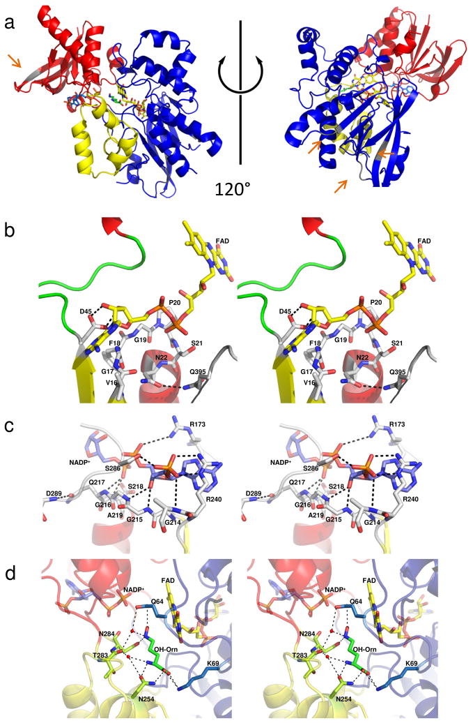
Figure 2.
Structure of the ornithine hydroxylase from Pseudomonas aeruginosa, PvdA. A) Cartoon of the oxidized structure of PvdA (PDB code 3S5W) [26]. The FAD binding domain is shown in blue, the NADPH binding domain in red and the ornithine binding domain in yellow. The FAD is yellow sticks, the NADP+ is blue sticks, and the ornithine is green sticks. The grey elements of secondary structure and loops regions represent areas of sequence insertion for SidA and are highlighted with orange arrows. B) Stereo image of the FAD binding site and C) Stereo image of the NADP+ binding site. In parts B and C, the cartoon is colored by secondary structure with α-helices red, β-strands yellow and loop regions green. D) Stereo image of the ornithine binding site. Here the cartoon is colored as in part A. Hydrogen bonding and salt links are shown as dashed lines.

