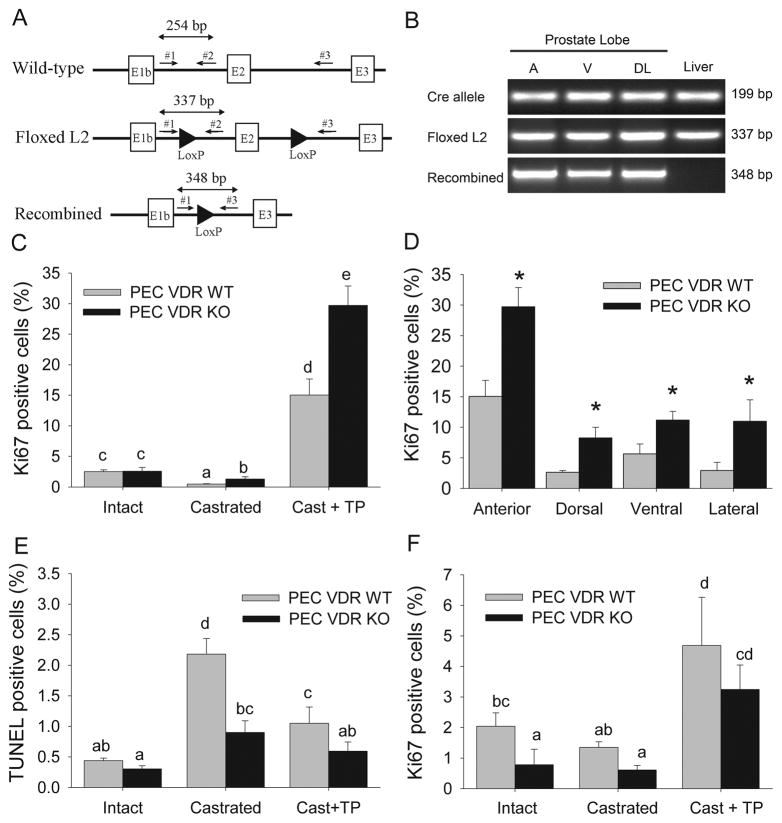
Figure 5. Prostate epithelial cell deletion of VDR regulates prostate cell proliferation and apoptosis.
Mice with prostate epithelial cell-specific deletion of the VDR (PEC VDR KO) or their wild-type littermates (PEC VDR WT) were subjected to castration (Cast) and testosterone repletion (Cast + TP). (A) Schematic of wild-type, floxed (L2), and Cre-recombined VDR alleles showing exons (boxes), LoxP sites (arrowheads) and PCR primers (arrows). (B) PCR analysis of VDR alleles. Cre-recombinase transgene = Cre allele, the floxed VDR L2 allele = Floxed L2, Cre-recombined VDR allele = Recombined. (C) Ki67 labeled prostate epithelial cells in the anterior lobe; (D) Ki67 labeled prostate epithelial cells in each of the lobes of the Cast + TP group; (E) TUNEL stained prostate epithelial cells in the anterior lobe; (F) Ki67 labeled prostate stromal cells in the anterior prostate. Bars are the mean±SE, n=6. In (C, E, F) bars without a common letter superscript are significantly different (p<0.05). In (D) * = significantly different from the PEC VDR WT group (p<0.05).

