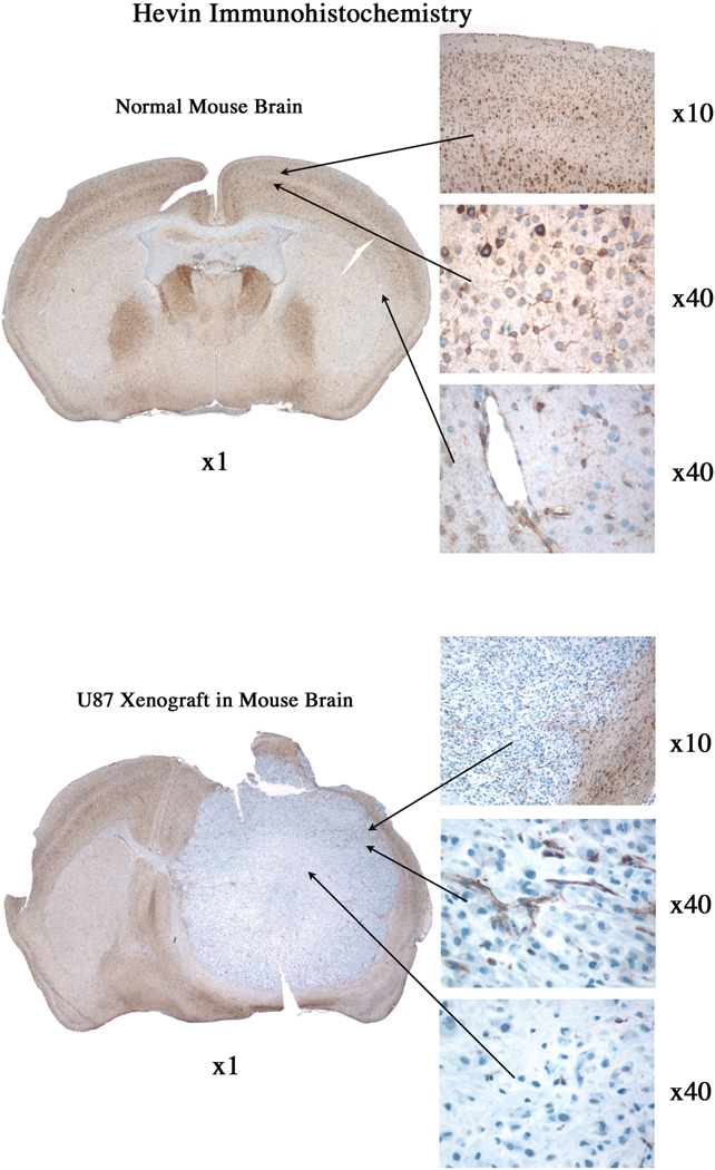
Figure 4. Hevin is expressed in normal mouse brain but not in U87 glioma cells.
5 µm sections of normal mouse brain (A) and mouse brain implanted with U87 glioma cells (B) were subjected to immunohistochemistry to detect hevin. A) Hevin was observed regionally in normal brain (×1 magnification), with high levels in neurons and astrocytes (×10, ×40 magnifications; top and middle panels) and blood vessels (×40 magnification; bottom panel). B) Hevin was undetectable in glioma U87 tumor cells (×1, ×40 magnification, bottom panel), but was present in surrounding normal brain (×10 magnification; top panel) and blood vessels (×40 magnification; middle panel).

