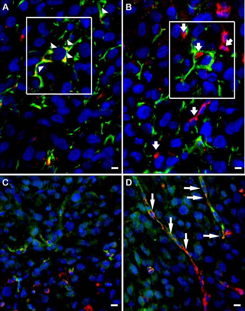
Figure 6. Hevin is located in glial infiltrates, whereas SPARC is found in microvasculature.
Gliomas that arose from A2b2 (SPARC-overexpressing) cells were subjected to immunohistochemistry with a combination of rabbit anti-GFAP IgG (A, B) or mouse anti-VWF IgG (C, D) (red), and goat anti-murine hevin IgG (A, C) or goat anti-murine SPARC IgG (green) (B, D). A) Co-localization of hevin (green) and GFAP (red) staining. B) Disparate localization of SPARC (green) and GFAP (red) staining. Boxed areas in A and B represent identical tumor locations in the serial sections. C) Disparate localization of hevin (green) and VWF (red). D) Co-localization of SPARC (green) and VWF (red). Arrowheads (A) indicate hevin+/GFAP+ cells. Short arrows (B) indicate SPARC (green) independent from GFAP (red). Long arrows (D) indicate SPARC+/VWF+ cells. Scale bars, 10 µm.

