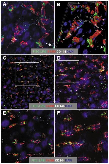
Figure 1. LPS remains within sinusoids for at least one week after i.v. injection.
Aoah−/− and Aoah+/+ mice were injected i.v. with 0.5μg FITC-LPS per g body weight. Liver sections were immunolabeled as in Methods to detect Kupffer cells (F4/80+, red), the junctions of sinusoidal endothelial cells (CD144 or VE-cadherin, white), and nuclei (blue). A. Aoah−/− liver, one day after injection (63x, zoom 2.4). B. 3-dimensional rendering of A. C and D. Images (x63, zoom 1) of Aoah+/+ (C) and Aoah−/− (D) livers on day 7 after injection. E and F. Higher magnification (x63, zoom 2.4) views of C and D (areas indicated by the rectangles). Seven days after injection, the LPS is seen largely within sinusoidal spaces and a large fraction of it is within, or closely associated with, Kupffer cells. Examples of extrasinusoidal LPS are indicated by the arrows in A and B.

