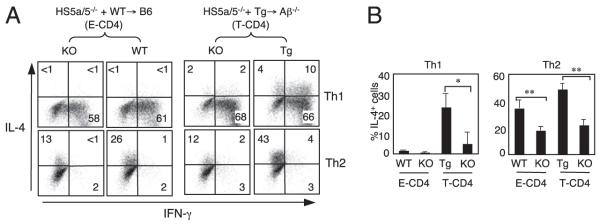
FIGURE 1.
The HS5 and HS5a region in the IL-4 locus is important but not essential to express IL-4. A and B, BM from HS5a/5−/− mice (knockout [KO]) (CD45.2) were mixed with those from WT or CIITA Tg mice (CD45.1/2) and cotransferred into B6 or Aβ−/− recipients (CD45.1). Eight to 12 wk after BM transplantations, splenic CD4 T cells from BM chimeras were differentiated into Th1 or Th2 cells and analyzed IL-4 and IFN-γ expression by intracellular cytokine staining. The cells were also stained with an anti-CD45.1 and anti-CD45.2 Ab to distinguish the two donors and the recipient cells. Graphs in B show the mean ± SD of four mice. *p < 0.05, **p < 0.01.

