FIGURE 2.
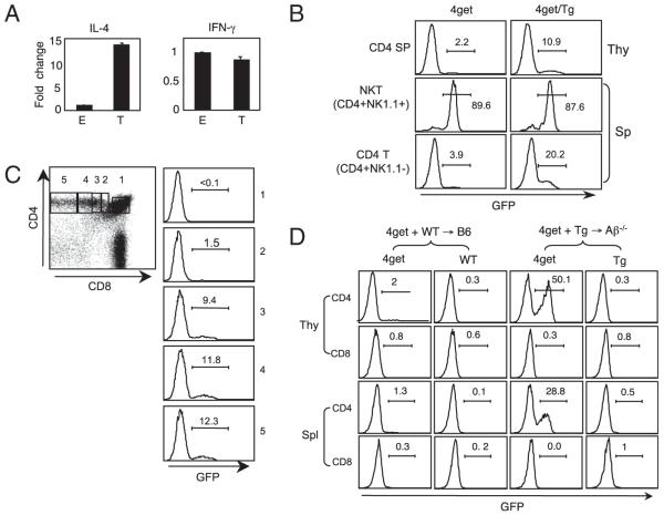
Expression of IL-4 during T-CD4 T cell development. A, IL-4 expression is enhanced in T-CD4 thymocytes. E- and T-CD4 T cells were electronically sorted from the thymus of [WT→B6] and [CIITATg→Aβ−/−] chimeric mice and subjected to RNA preparation immediately. IL-4 and IFN-γ mRNA were quantified by quantitative RT-PCR, and the results were expressed as ratios relative to the housekeeping gene GAPDH. B, GFP expression of 4get and 4get mice expressing CIITA transgene (4get/Tg). Thymocytes and splenocytes from 4get and 4get/Tg mice were freshly isolated and used to assess GFP expression. Numbers indicate the percentage of cells expressing GFP. Representative profiles are shown. C, GFP expression during CD4 T cell development in the 4get/Tg thymus. Total thymocytes were used to measure GFP expression. Numbers in the dot plot correspond to each histogram. D, Thymic selection dictates IL-4 expression measured by GFP. Chimeric mice [4get+WT→B6] and [4get+Tg→Ab−/−] were generated to obtain E- and T-CD4 T cells, respectively. Eight to 12 wk after BM transplantations, total thymocytes and splenocytes were prepared and examined for GFP expression without manipulation in vitro. The numbers shown are the percentage of GFP+ cells. A–C, Data are representative of more than three independent experiments; (D) data are representative of two independent experiments.

