FIGURE 3.
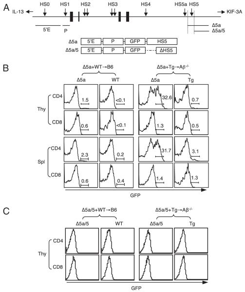
HS5 together with the 5′ enhancer and the promoter can induce IL-4 expression in T-CD4 T cells. A, Schematic diagram of the IL-4 locus and the transgene constructs used in this study. Black boxes denote IL-4 exons; arrows denote HS sites; horizontal bars represent fragments used for Tg constructs. B, HS5-mediated expression of GFP in T-CD4 T cells. BM from Δ5a mice (CD45.2) shown in A were mixed with WT or Tg BM (CD45.1/2) and cotransferred to either B6 or Aβ−/− hosts (CD45.1). Chimeric mice were reconstituted for 8–12 wk. Freshly isolated total thymocytes and splenocytes were used to assess GFP expression by flow cytometry. The numbers shown are the percentage of GFP+ cells. C, Lack of GFP expression in the absence of HS5. Mixed BM chimeras were constructed as in A except BM cells from Δ5a/5 reporter mice shown in A were used. GFP expression of CD4 and CD8 T cells in the thymus is shown.

