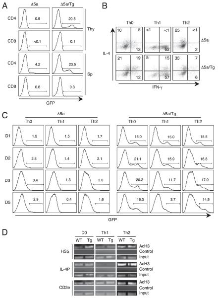
FIGURE 6.
Loss of HS5-mediated IL-4 expression by the Th1-inducing signal. A, Total thymocytes (Thy) or splenocytes (Sp) from HS5 mice (Δ5a) shown in Fig. 3A or HS5 crossed with CIITATg mice (Δ5a/Tg) were compared for GFP expression. B, Splenic CD4 T cells were differentiated under non-skewing (Th0), Th1, or Th2 conditions from Δ5a and Δ5a/Tg mice as shown in Fig. 3A and were analyzed for IL-4 and IFN-γ. C, Splenic CD4 T cells from Δ5a mice or Δ5a/Tg mice were differentiated as in B and analyzed for GFP expression at the indicated time points. Numbers indicate the percentage of GFP+ cells. D, Differential acetylation of histone H3 at HS5. WT and CIITA Tg mice were used to isolate splenic CD4 T cells. Freshly isolated (D0) or cells cultured for 5 d under the Th1 and Th2 differentiation were subjected to ChIP assays as in the Materials and Methods followed by PCR. The same precipitated DNA was used for PCR to assess the level of acetylation at HS5, IL-4 promoter (IL-4P), and CD3ε. No Ab group was used as a negative control. CD3ε and input DNA was used as an internal control. Data shown are representative from three independent experiments.

