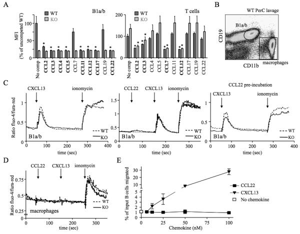
Figure 6. Chemokine binding and signalling properties of D6 on PerC B1 cells.

(A) CCL2AF647 uptake by WT and D6-deficient (KO) PerC B1a/b (CD19+CD11b+) or T cells (CD19−CD11b−CD5+) in the presence of a 10-fold molar excess of the indicated unlabeled chemokines. ‘No comp’; CCL2AF647 uptake in the absence of competitor. The average Mean Fluorescence Intensity (MFI) of competed samples is shown as percent (+/−SEM) of the average MFI of uncompeted WT uptake. Asterisks mark chemokines that significantly reduced CCL2AF647 uptake by WT or KO cells (p<0.01). (B) Gates used to gauge Ca2+ fluxes in B1a/b and MOs. (C-D) Representative Ca2+ flux profiles of Fluo-4/Fura-Red-loaded WT and KO PerC B1a/b cells (C) and MOs (D). The points of chemokine (50-100nM) and ionomycin addition are indicated with arrows. Cells in the far right panel of C were incubated in 100nM CCL22 for 1h at 37°C before analysis. (E) Migration of WT B cells from PerC lavages to CCL22 or CXCL13. Migration in media alone is indicated by the unfilled square. The mean number of live cells (+/−SEM) migrated from 4 replicate wells is presented as percent of input. All data are representative of 3 or more independent experiments.
