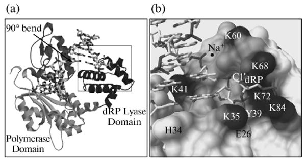
Fig. 5.

Global structure of polymerase β binary complex bound to DNA. (a) The conformation of the polymerase β binary complex bound to dRP-containing DNA (sugar ring in closed form) is shown. The amino-terminal 8-kDa lyase domain and the polymerase sub-domain are indicated. The structure is identical to that observed previously with gapped DNA [41], except that the downstream primer contains a 5′-THF phosphate (dRP, in boxed area). An arrow indicates the position of a 90° bend in the template strand. DNA is shown as a stick model. (b) A close-up view of the boxed area in (a).
