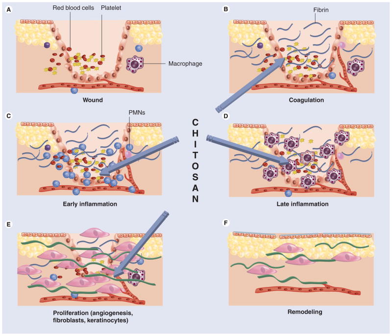
Figure 4. The phases of cutaneous wound healing.
(A) Immediately following cutaneous injury, blood elements and amines extravasate from locally damaged blood vessels within the dermis. Vascular permeability is temporarily increased to allow neutrophils (PMNs), platelets and plasma proteins to infiltrate the wound. Vasoconstriction follows, in response to factors released by these cells. (B) Coagulation then occurs as platelets aggregate with fibrin, which is deposited in the wound following its conversion from fibrinogen. (C) Platelets release several factors, including PDGF and TGF-β, which attract PMNs to the wound, signaling the beginning of inflammation. (D) After 48 h, macrophages replace PMNs as the principal inflammatory cell. Together, PMNs and macrophages remove debris from the wound, release growth factors and begin to reorganize the extracellular matrix. (E) The proliferation phase begins at about 72 h as fibroblasts, recruited to the wound by growth factors released by inflammatory cells, begin to synthesize collagen, and angiogenesis and re-epitheliazation occurs. (F) Collagen crosslinking and reorganization occur for months after injury in the remodeling phase of repair. Chitosan has been reported to beneficially influence stages (B–E).
PMN: Polymorphonuclear leukocyte.
Reproduced with permission from [108].

