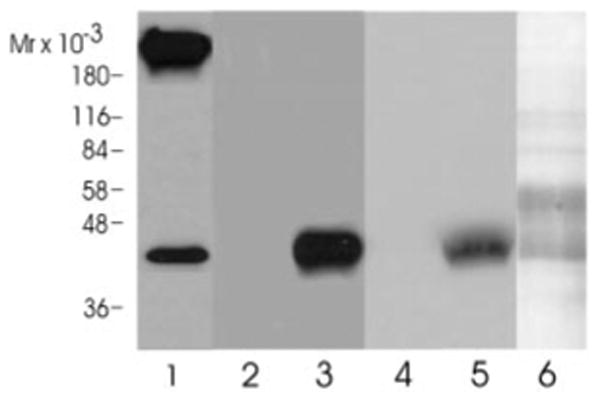
Fig. 8. Western blot analysis.

Aliquots of the crude membrane fraction (lane 1), the α-cobratoxin eluate (lanes 2 and 3), and the lentil lectin column eluate (lanes 4 and 5) were analyzed by SDS-gel electrophoresis and immunoblotted onto a polyvinylidene difluoride membrane. Blots were probed either with the preimmunserum (lanes 2 and 4) or the affinity purified Dα5 antibody (1:500 dilution). Bound antibodies were visualized with a horseradish peroxidase-conjugated anti-rabbit antibody (Bio-Rad) using an enhanced chemiluminescence Western blot kit (Pierce). The antibody detected a protein of 42 kDa in lanes 1, 3, and 5. Lane 6 shows a silver stain of the purified receptor fraction that was run on the gel in parallel to the samples in lanes 4 and 5.
