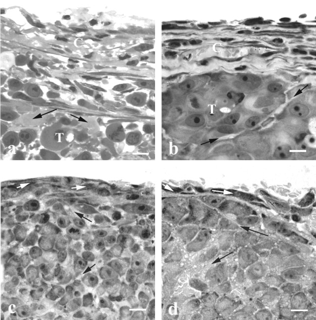
Figure 2.

Light microscopy (LR White sections) of the peripheral region of DC and CW tumors. The capsule of U87 (a) and Mu89 (b) DC tumors is composed of several layers of fibroblast-like cells separated by ECM. Note the large intercellular spaces in U87 and the narrow space that separates two cellular nodules in Mu89. The connective tissue at the edge of U87 (c) and Mu89 (d) in the CW is composed of one fibroblast cell layer; the tumor cells are separated by narrow intercellular spaces. C, capsule; T, tumor; black arrows, ECM; white arrows, fibroblast-like cells. (Bar = 10 μm.)
