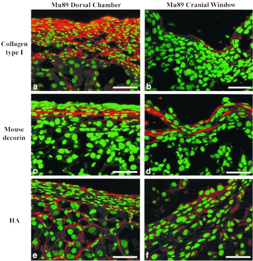
Figure 3.
Immunostaining for collagen type I (a and b) and decorin (c and d), and labeling for HA (e and f) in DC (a, c, and e) and CW (b, d, and f) tumors. Collagen type I occupies a greater area of the periphery in DC than in CW tumors. In both DC and CW tumors the decorin staining is restricted to the periphery of the tumor. HA staining is intense in the center of Mu89 in the DC, whereas in the periphery the staining is weak. (Bar = 100 μm.)

