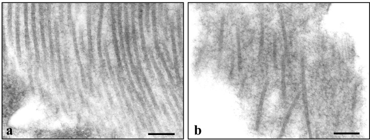
Figure 4.

Electron microscopy of the organization of collagen fibrils in the capsule of U87 tumors in the DC. (a) The longitudinally oriented fibrils are parallel to one another with an interfibrillar spacing that varies from 20 to 42 nm. (b) The fibrils are poorly organized. The interfibrillar spacing varies between 75 and 130 nm. (Bar = 200 nm.)
