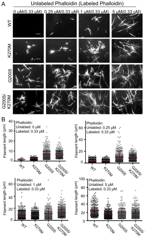
Figure 7. Substituted TgACTI alleles demonstrate enhanced in vitro polymerization.
(A) In vitro polymerization of recombinant TgACTI alleles substituted with mammalian actin residues. Actins (5 µM) were visualized by fluorescence microscopy using 0.33 µM Alexa 488-phalloidin and different molar ratios of unlabeled phalloidin to actin. Scale bars, 5 µm. Representative of three or more similar experiments. The values at the top of the figure panel indicate the amounts of phalloidin used: µM unlabeled phalloidin (µM labeled phalloidin). (B) Quantitation of filaments formed during in vitro polymerization of substituted TgACTI alleles. Mean ± S.D. shown by horizontal line. Concentrations of phalloidin used for treatment are indicated below the graphs as µM unlabeled phalloidin (µM labeled phalloidin). In the presence of only low levels of labeled phalloidin (i.e. 0.33 µM), filaments formed by TgACTI-K270M (P<0.01), TgACTI-G200S (P<0.001), and TgACTI-G200S/K270M (P<0.001) were significantly longer that wild type TgACTI (Student's t-test).

